← Back to Unit Converters

Select Radiation Quantity

This converter works within each category (activity, exposure, absorbed dose, equivalent dose). Cross-category questions like “Bq to Gy” require extra physical context – see the explanation in the Formula section.

Enter Value & Units

Result

Converted:

Scientific:

Grouped:


⏳ Radioactivity Decay Calculator (Half-Life)

Compute how activity changes over time using the exponential decay law. Units for activity (Bq, kBq, MBq, Ci) are handled by the same multiplier.

Decay Result

Remaining activity A(t):

Fraction remaining:

Fraction decayed:

📏 MU ↔ Dose Helper (Machine-Specific)

For linear accelerators calibrated at reference conditions, dose is often approximately proportional to monitor units (MU). Enter your own calibration – this helper does not replace treatment planning.

Monitor Units: –

⚠️ Clinical safety: Always verify MU with your department’s TPS / QA procedures. This helper is for educational use only.

🔍

    ☢️ Overview: Radiation Unit Converter & Dose Context

    This radiation unit converter is designed to help you move quickly between the most common radiation units used in medical imaging, nuclear medicine, radiation therapy and radiation protection. It focuses on four distinct physical quantities – each with its own unit family:

    • Source activity – how many radioactive decays happen per second.
      • Becquerel (Bq) – SI unit, 1 Bq = 1 decay per second.
      • Curie (Ci) – legacy unit, 1 Ci = 3.7×10¹⁰ Bq.
      • Rutherford (Rd) – rarely used; 1 Rd = 106 decays/s.
      • Disintegrations per second (1/s) – physically identical to Bq.
    • Exposure in air – ionisation produced in air.
      • Roentgen (R)
      • Coulomb per kilogram (C/kg) – SI unit for exposure.
    • Absorbed dose – energy deposited per kilogram of material.
      • Gray (Gy) – SI unit, 1 Gy = 1 joule/kg.
      • Rad – older unit, 1 rad = 0.01 Gy.
      • Joule per kilogram (J/kg) – equivalent to Gy.
    • Equivalent dose – absorbed dose weighted for biological effect.
      • Sievert (Sv) – SI unit used for risk and dose limits.
      • Rem – older unit, 1 rem = 0.01 Sv.

    The calculator converts within each group (for example Gy ↔ rad, Sv ↔ rem, Bq ↔ Ci), but does not perform cross-category conversions such as “Bq to Gy” or “R to Sv” – those require additional assumptions about the source, geometry, shielding and tissue. This separation keeps the results physically meaningful and avoids misleading shortcuts.

    Below the core converter, you will also find:

    • A radioactivity decay (half-life) calculator to estimate how activity changes over time.
    • A simple MU ↔ dose helper for linear accelerators, allowing you to relate monitor units to planned dose using your own calibration. Monitor units (MU) are the internal “output counts” of a linac and are typically calibrated so that a certain number of MU delivers a known dose, such as 1 cGy per MU at reference conditions.

    This page also includes a radiation dose comparison table and dose examples so you can put your converted values into context – for example, how a CT scan compares with background radiation or a chest X-ray.

    📈 Radiation Dose & Units – Visual Comparison (Infographic)

    Infographic showing radiation unit families Bq, Gy, Sv, rad, rem, R and typical effective doses from medical imaging compared to natural background radiation.
    This visual guide summarizes the main radiation unit families (source activity, exposure, absorbed dose, equivalent / effective dose) and compares typical effective doses from everyday exposures and medical imaging (banana equivalent dose, airport scan, dental X-ray, chest X-ray, CT scans, space missions) to natural background radiation. Values are approximate educational examples based on published sources such as FDA guidance and ICRP / UNSCEAR reports.

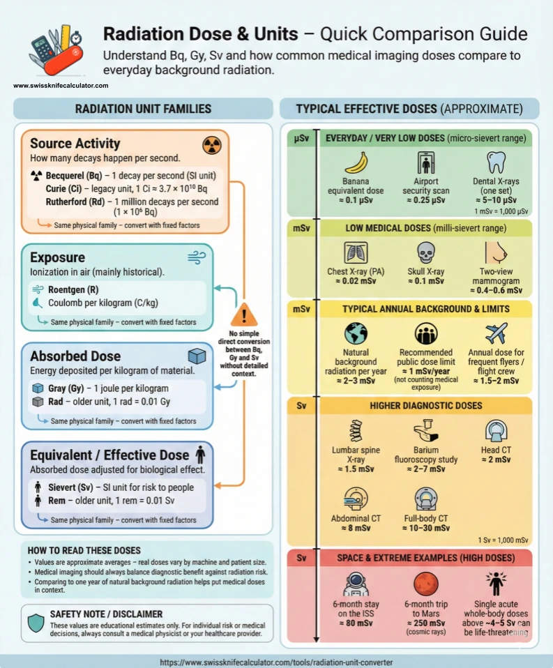
    Use this infographic together with the Radiation Unit Converter results: after you convert a dose in mGy or mSv, you can quickly place it on the scale – for example, “this dose is similar to a chest X-ray” or “this is in the range of a single CT scan” – and compare it to annual background radiation.

    🧮 Formula & Methodology: How the Radiation Converter Works

    For each radiation quantity, the converter uses a simple two-step method with SI base units in the middle:

    Step 1 – Convert input to SI base unit
      value_SI = value_input × factor_from
    
    Step 2 – Convert SI value to target unit
      value_target = value_SI ÷ factor_to

    The SI base unit is: Bq for activity, C/kg for exposure, Gy for absorbed dose and Sv for equivalent dose.

    Key conversion relationships used by the tool

    Activity

    1 Ci  = 3.7 × 10^10 Bq
    1 Rd  = 1 × 10^6 decays/s
    1 Bq  = 1 decay/s = 1 s^-1

    Exposure in air

    1 R   ≈ 2.58 × 10^-4 C/kg

    Absorbed dose

    1 Gy  = 1 J/kg
    1 rad = 0.01 Gy
    1 Gy  = 100 rad

    Equivalent dose

    1 Sv   = 100 rem
    1 rem  = 0.01 Sv

    Half-life & radioactivity decay

    The decay calculator uses the standard exponential decay law. Starting from an initial activity A0 with half-life T1/2 and elapsed time t, the remaining activity A(t) is given by:

    A(t) = A0 × (1/2)^(t / T1/2)
    
    or equivalently
    
    λ      = ln(2) / T1/2
    A(t)   = A0 × e^(−λ × t)

    The tool automatically converts the half-life and elapsed time into seconds (from seconds, minutes, hours, days or years) so the formula is applied consistently regardless of the units you choose.

    MU ↔ dose helper (monitor units)

    In external beam radiotherapy, a linear accelerator (linac) delivers dose in terms of monitor units (MU). MU is a dimensionless counter that reflects how long and under which conditions the beam is on. At reference conditions (field size, SSD/SSD, depth, beam energy), the machine is calibrated so that each MU corresponds to a known dose, often:

    Calibration example:
      1 MU → 1 cGy at reference point in water

    In this simplified situation, dose and MU are roughly proportional. If your calibration is k cGy per MU and you plan a fraction dose of D_Gy in gray, the approximate MU is:

    1 Gy  = 100 cGy
    
    Given:
      k      = calibration in cGy/MU
      D_Gy   = planned dose in Gy
    
    Convert dose to cGy:
      D_cGy  = D_Gy × 100
    
    Compute MU:
      MU ≈ D_cGy ÷ k

    For example, with k = 1.0 cGy/MU and D_Gy = 2.0 Gy:

    D_cGy = 2.0 × 100 = 200 cGy
    MU    = 200 cGy ÷ 1.0 cGy/MU = 200 MU

    In real clinical practice, additional factors (field size, depth, beam modifiers, SSD/FS, off-axis ratios, dynamic techniques, heterogeneities) make the relationship between dose and MU more complex. This helper shows the basic proportional idea for education; clinical MU must always come from a verified treatment planning system and QA process.

    📊 Worked Examples Using the Converter

    1. Activity: Curie → Becquerel

    Imagine a PET isotope with an activity of 1 Ci. In the Source Activity mode:

    • Enter 1 as the value.
    • Select Curie (Ci) as “From”.
    • Select Becquerel (Bq) as “To”.

    The converter uses:

    1 Ci × (3.7 × 10^10 Bq / Ci) = 3.7 × 10^10 Bq

    You will see a converted value of about 37,000,000,000 Bq (3.7000e+10 Bq).

    2. Absorbed dose: rad → gray

    You have an older report quoting 100 rad. In the Absorbed Dose mode:

    • Value = 100
    • From = Rad (rad)
    • To = Gray (Gy)

    The converter uses:

    1 rad = 0.01 Gy
    
    100 rad × 0.01 Gy/rad = 1 Gy

    Result: 1 Gy.

    3. Equivalent dose: Sv → rem

    Suppose an occupational exposure is 0.05 Sv (which equals 50 mSv). In the Equivalent Dose mode:

    • Value = 0.05
    • From = Sievert (Sv)
    • To = Rem (rem)

    The converter uses:

    1 Sv = 100 rem
    
    0.05 Sv × 100 rem/Sv = 5 rem

    Result: 5 rem.

    4. Exposure: R → C/kg

    For an old survey meter reading of 0.2 R, in the Exposure mode:

    • Value = 0.2
    • From = Roentgen (R)
    • To = Coulomb per kilogram (C/kg)

    The calculator uses:

    1 R ≈ 2.58 × 10^-4 C/kg
    
    0.2 R × (2.58 × 10^-4 C/kg per R)
      = 5.16 × 10^-5 C/kg

    The tool shows approximately 5.16×10⁻⁵ C/kg.

    5. Half-life example

    A radionuclide has a half-life of 6 days and an initial activity A0 = 1000 (for the fraction, the absolute unit cancels). After 12 days:

    • Half-life = 6, unit = days
    • Elapsed time = 12, unit = days

    The mathematics behind the result is:

    Number of half-lives = t / T1/2 = 12 / 6 = 2
    
    A(t) = A0 × (1/2)^(2)
         = 1000 × (1/4)
         = 250
    
    Fraction remaining = 250 / 1000 = 0.25
    Fraction decayed   = 1 − 0.25 = 0.75 (75%)

    The decay calculator will report about 250 remaining with a fraction remaining of 0.25 and fraction decayed of 0.75.

    6. MU helper example

    If your linac is calibrated to 1.0 cGy/MU at reference and you plan a fraction dose of 2.0 Gy:

    Dose in cGy:
      D_cGy = 2.0 Gy × 100 cGy/Gy = 200 cGy
    
    MU with k = 1.0 cGy/MU:
      MU = D_cGy ÷ k
         = 200 cGy ÷ (1.0 cGy/MU)
         = 200 MU

    The helper will display roughly 200 MU, which matches the intuitive idea that — in this simple calibration — 100 MU ≈ 1 Gy at reference conditions.

    📈 Visual Guide: Typical Diagnostic Doses & Background Radiation

    Converting units is only half of the story. To interpret a result, it helps to compare it with typical medical procedures and everyday background radiation. The table below summarises approximate effective doses for common exams and how they relate to a chest X-ray and to natural background (assuming about 3 mSv per year from background radiation).

    Approximate effective dose comparison for common X-ray & CT procedures
    Diagnostic procedure Typical effective dose (mSv) ≈ chest X-rays (PA) * ≈ natural background time **
    Chest X-ray (PA film) 0.02 1 ≈ 2.4 days
    Skull X-ray 0.1 ≈ 5 ≈ 12 days
    Lumbar spine X-ray 1.5 ≈ 75 ≈ 6 months
    I.V. urogram 3 ≈ 150 ≈ 1 year
    Upper GI exam 6 ≈ 300 ≈ 2 years
    Barium enema 8 ≈ 400 ≈ 2.7 years
    CT head 2 ≈ 100 ≈ 8 months
    CT abdomen 8 ≈ 400 ≈ 2.7 years

    * Chest X-ray comparison assumes ~0.02 mSv per PA film. ** Background radiation comparison assumes ~3 mSv per year. Values are rounded and represent typical ranges; individual exams can be higher or lower depending on patient size and protocol.

    Real-world dose examples

    The following approximate figures are commonly cited in radiation protection literature and help you build intuition about the numbers produced by the converter:

    • ~0.1 µSv – “banana equivalent dose”, a tongue-in-cheek unit for a single banana.
    • ~0.25 µSv – typical limit for one airport security scan.
    • 5–10 µSv – one set of dental X-rays.
    • 0.4–0.6 mSv – two-view mammography.
    • ~1 mSv per year – recommended public dose limit in many regulations (excluding medical and occupational exposure).
    • 2.4 mSv per year – global average natural background dose.
    • 10–30 mSv – a single full-body CT scan, depending on protocol.
    • 50 mSv per year – typical upper occupational dose limit in many standards.
    • hundreds of mSv – doses received by some nuclear workers and accident responders.
    • several Sv (sievert) – range associated with serious radiation sickness and potentially fatal acute exposure.

    How to interpret your own result safely

    • Keep units consistent. If your report says “10 mSv” and the converter uses Sv, enter it as 0.01 Sv.
    • Compare, don’t self-diagnose. Use the table and examples to understand the order of magnitude of a dose, but always discuss risk and benefits with your doctor or medical physicist.
    • Single exam vs. lifetime. Many diagnostic procedures deliver a small fraction of annual background radiation or occupational limits. Risk depends on cumulative exposure and individual health.
    • Context matters. 5 mSv in a one-off CT scan is not the same situation as 5 mSv every month for many years.

    For quick scientific formatting of very large or very small numbers, you can also use the Scientific Notation Converter available on SwissKnifeCalculator.

    🔬 Practical Use Cases for This Radiation Converter

    • 🩻 Radiology & imaging reports – convert between Gy, rad, Sv and rem when comparing older literature with modern SI-based reports.
    • ⚛️ Nuclear medicine – translate activity values from Ci to Bq when working with dose calibrators, PET tracers and regulatory paperwork.
    • 💼 Occupational dose tracking – convert between Sv and rem or between mSv and Sv to compare personal dosimeter readings with dose limits.
    • 🎓 Teaching & exam preparation – quickly check homework exercises and lab problems involving activity, dose and exponential decay.
    • 🏥 Patient information – prepare understandable comparisons (for example “this CT scan is about X years of background radiation”) while keeping units consistent.
    • 🧪 Research & QA – sanity-check exposure and dose calculations from spreadsheets, Monte-Carlo simulations or treatment planning exports.
    • 🎯 Radiotherapy education – use the MU helper to understand how Gy, cGy and MU are related at reference conditions, and why additional factors are needed in real treatment planning.

    For more engineering-style tools, you can also explore the Energy & Environment tools and other unit converters on SwissKnifeCalculator.

    ❓ Frequently Asked Questions About Radiation Units

    Why does the converter separate Bq, Gy and Sv into different modes?

    Bq measures activity (decays per second), Gy measures absorbed dose (energy per kilogram), and Sv measures equivalent dose (dose weighted by biological effect). They describe different physical quantities, so there is no universal “Bq to Gy” or “Gy to Sv” factor. The converter therefore keeps them in separate modes and only converts between units that measure the same thing.

    Can I convert Bq to Gy or Gy to Sv with this tool?

    Not directly. To go from activity to dose you would need detailed information about the radionuclide, emission energy, geometry, shielding, medium and exposure time. Even in professional work, this is normally handled by dose-conversion coefficients, Monte-Carlo simulations or treatment planning systems – not by a simple unit converter.

    How do I enter mSv or µSv if the dropdown only lists Sv?

    Use decimal values. For example:

    • 2 mSv = 0.002 Sv
    • 500 µSv = 0.0005 Sv

    The converter then treats the value correctly in Sv and you can mentally convert back to mSv or µSv if you prefer.

    What is the difference between gray (Gy) and sievert (Sv)?

    Gy tells you how much energy per kilogram was absorbed, regardless of radiation type. Sv includes a radiation weighting factor to reflect biological effect. For X-rays and gamma rays, 1 Gy often corresponds roughly to 1 Sv, but for neutrons or alpha particles the same Gy can represent several Sv.

    Is rem still used, or should I always use Sv?

    In modern scientific and international regulatory documents, Sv is the standard. However, rem is still found in older reports, U.S. regulations and legacy dosimetry systems. The converter makes it easy to translate: 1 Sv = 100 rem and 1 rem = 0.01 Sv.

    What exactly is a monitor unit (MU) in radiotherapy?

    A monitor unit (MU) is the linac’s internal “output counter”. It is dimensionless and is defined by your department’s calibration protocol. Typically, the machine is set up so that a certain number of MU delivers a known dose (for example 1 MU = 1 cGy at a specific depth and field size in water). Once that calibration is fixed, changing MU changes the delivered dose in a roughly proportional way, while other factors adjust for depth, field size, dynamic techniques, and so on.

    Can I use the MU helper for real patient treatments?

    No. The MU helper on this page is a simplified educational calculator. Real clinical treatments must be planned and verified with your department’s treatment planning system (TPS), independent checks and QA protocols. The helper is useful to understand the rough relationship between dose, cGy and MU, but it is not a treatment planning tool.

    How accurate are the “typical dose” values in the comparison table?

    They are order-of-magnitude averages compiled from published radiology and radiation protection references. Actual dose for an individual scan can vary significantly depending on scanner type, protocol, patient size and modern dose-reduction techniques. Always consult your radiologist or medical physicist for exam-specific values.

    Is a CT scan dose dangerous compared with background radiation?

    A single CT scan typically delivers a dose in the range of a few to a few dozen millisieverts – similar to several months to a few years of natural background radiation. Whether that is medically justified depends on the diagnostic benefit, your health situation and cumulative exposure over time. These decisions should always be made together with your physician, not based on an online calculator alone.

    What annual dose limits are commonly used for workers and the public?

    Many regulations adopt limits broadly in line with ICRP recommendations, such as:

    • 1 mSv per year above background for the general public.
    • Up to 20–50 mSv per year averaged over several years for radiation workers.

    Exact limits vary by country, profession and age group, so always check your local regulations and workplace rules.

    How should I cite or reference numbers from this page?

    If you are writing a report, assignment or presentation, you should reference primary sources such as radiology dose catalogues, ICRP publications, regulatory guidance documents or textbooks. You can use this tool to check unit conversions and orders of magnitude, then look up the exact values in the original documents.

    ⚠️ Important Disclaimer

    This radiation unit converter is provided for educational and informational purposes only. It:

    • does not calculate patient-specific risk or diagnose disease,
    • does not replace clinical judgement, treatment planning or regulatory advice,
    • uses typical reference values that may not match your local equipment or protocols.

    Never delay or refuse a medically indicated imaging or treatment procedure because of information from this page. Always discuss questions about your personal radiation exposure with your doctor, radiologist or medical physicist.

    Reviewed: December 2025. Values and examples are approximate and may be updated as new reference data become available.

    Data quality: Radiation dose values on this page are based on peer-reviewed medical physics literature, international radiation-protection guidelines (ICRP, UNSCEAR) and clinical imaging dose tables. They are intended for education and risk comparison, not for individual medical decision-making.

    📚 Sources & Further Reading

    1. Mettler FA Jr, Huda W, Yoshizumi TT, Mahesh M. Effective Doses in Radiology and Diagnostic Nuclear Medicine: A Catalog. Radiology. 2008;248(1):254–263.
    2. International Commission on Radiological Protection (ICRP). The 2007 Recommendations of the International Commission on Radiological Protection. ICRP Publication 103.
    3. United Nations Scientific Committee on the Effects of Atomic Radiation (UNSCEAR). Sources and Effects of Ionizing Radiation. UNSCEAR 2000 & 2008 Reports.
    4. CT scan – typical effective dose table (accessed 2025).
    5. Harvard Health Publishing – Radiation risk from medical imaging (overview of imaging dose ranges).
    6. Sievert – dose and dose-rate examples (summarises many of the “dose examples” and “dose rate examples” used above).

    The dose and dose-rate values on this page are approximate, rounded ranges adapted from these references. Actual patient dose depends on equipment, protocol and patient size.